MEDIDENT · CURRICULUM & CLINICAL INFORMATION
Radiodiagnosis and Medical Imaging
Radiographic techniques, imaging and interpretation relevant to teeth, jaws, skull and the orofacial region.
Related dental speciality: Oral and Maxillofacial Radiology
Body regions covered in radiodiagnosis and medical imaging
Teeth and supporting structures; jaws and maxillofacial bones; the skull; temporomandibular joint and salivary-region structures where covered by the cited imaging curriculum; and the head and neck region. The original curriculum screenshots below show maxillofacial radiology, radiodiagnostic techniques, radiological anatomy and interpretation.
Evidence: See the original DCI curriculum screenshots and explanations directly below. Each screenshot is retained as supplied. The curriculum level and individual practitioner’s qualifications, competence and applicable regulations determine professional scope.
Curriculum evidence · Imaging
Maxillofacial Radiology, Radiodiagnosis and Diagnostic Imaging Techniques
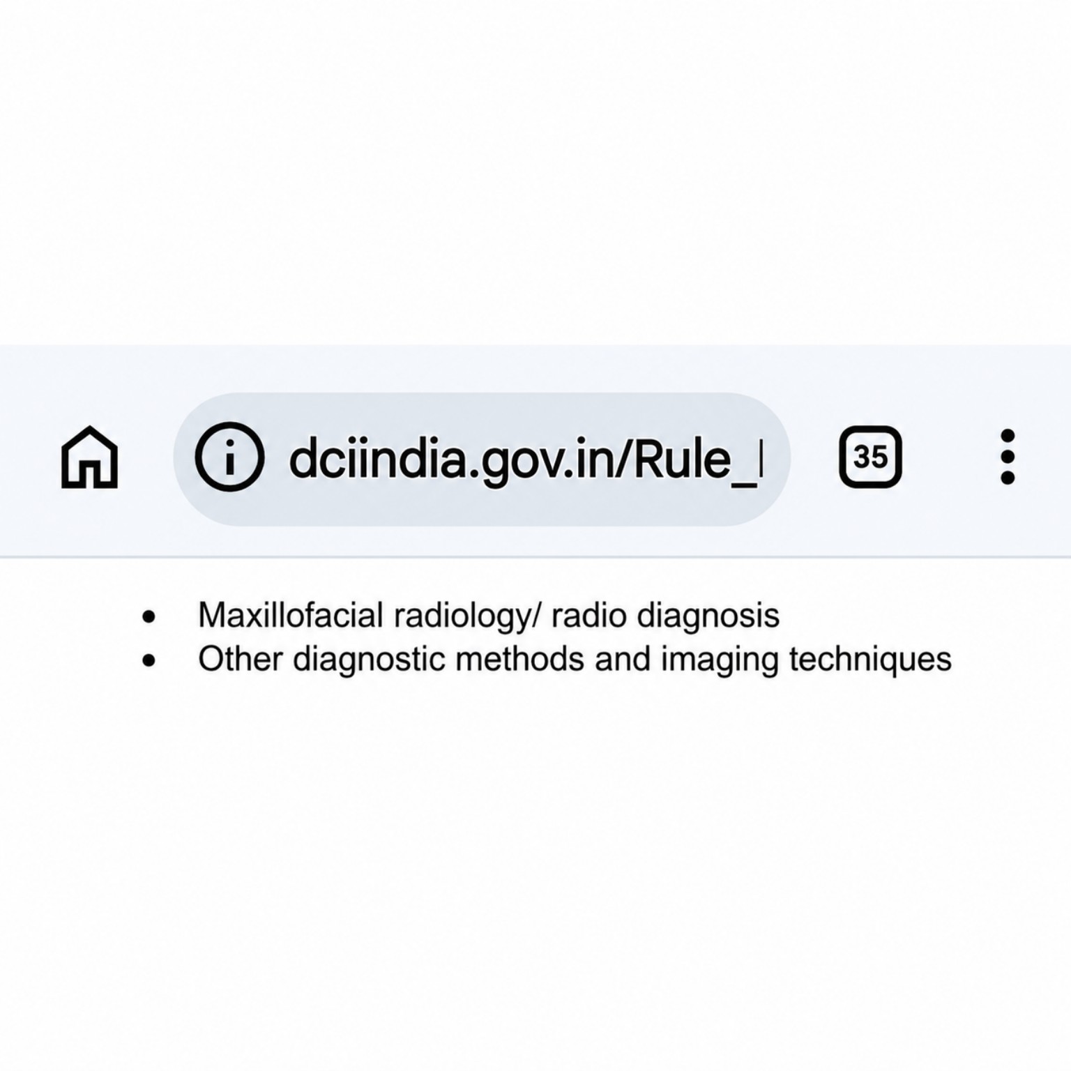
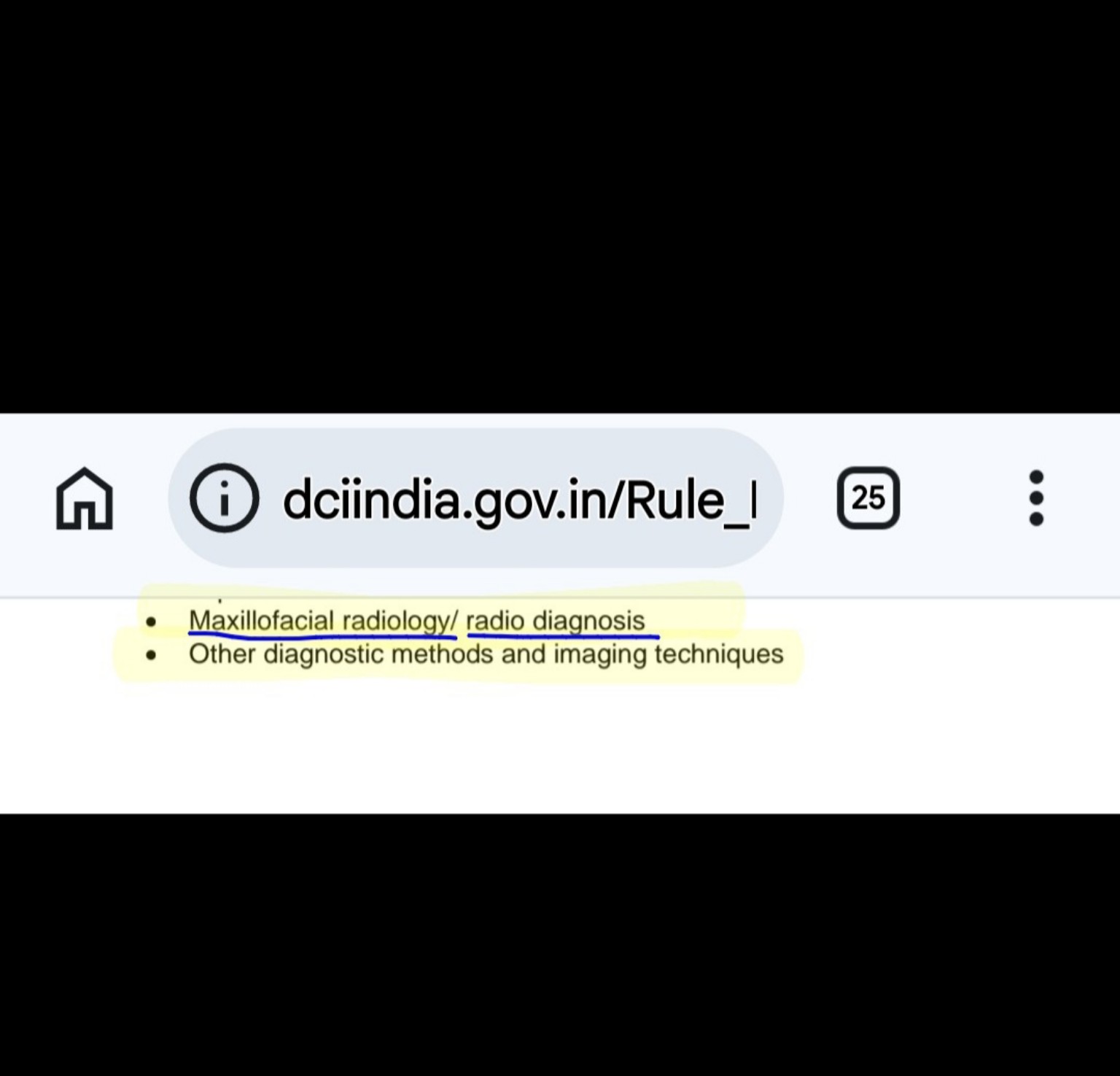
The DCI curriculum lists Maxillofacial radiology/radio diagnosis and other diagnostic methods and imaging techniques. This is curriculum evidence of training that includes maxillofacial radiological diagnosis and diagnostic imaging methods.
Original DCI curriculum screenshot – Curriculum evidence
Curriculum evidence · Imaging
Radiodiagnosis of the Head and Neck Region
The DCI MDS Radiology syllabus specifically lists radiological techniques of the head and neck region, radiological anatomy of the head and neck, interpretation of X-ray films and diagnostic applications. The accompanying clinical, practical and viva-voce examination description includes case studies, diagnosis and treatment planning for oral diseases, radiological techniques and radiological diagnosis.
Original DCI curriculum screenshot – Curriculum evidence
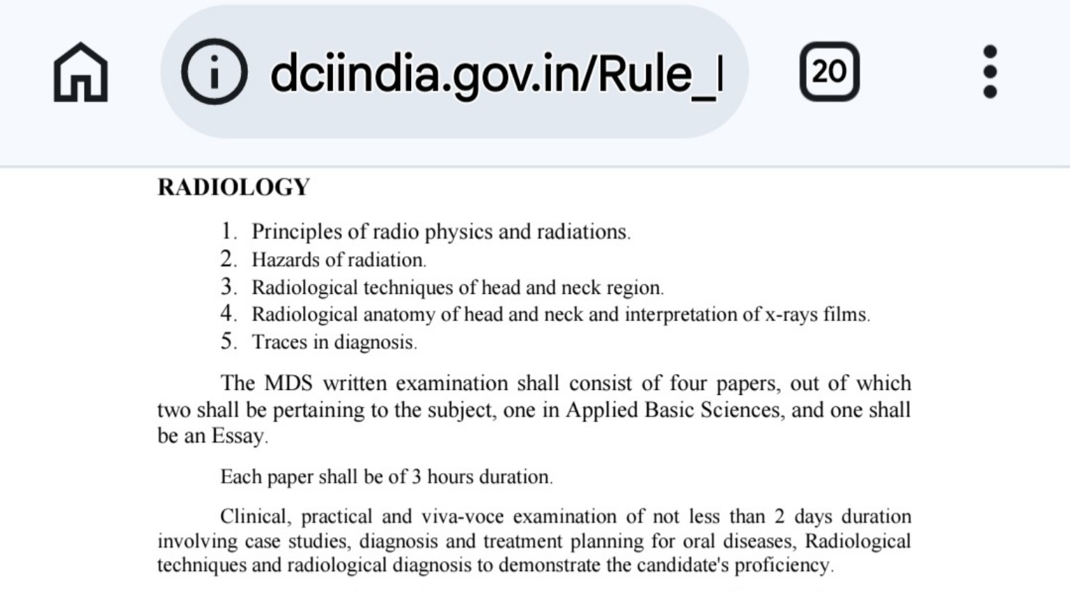
Scope clarification: This screenshot is from the MDS syllabus. It documents the stated Radiology training and assessment in that postgraduate curriculum; it should not be presented as proof that every BDS graduate has the same level of postgraduate radiology training.
Official source: Dental Council of India – MDS Course Regulations, Radiology syllabus.
Back to all evidenceCurriculum evidence · Imaging
Radiodiagnosis of Orofacial Region Problems
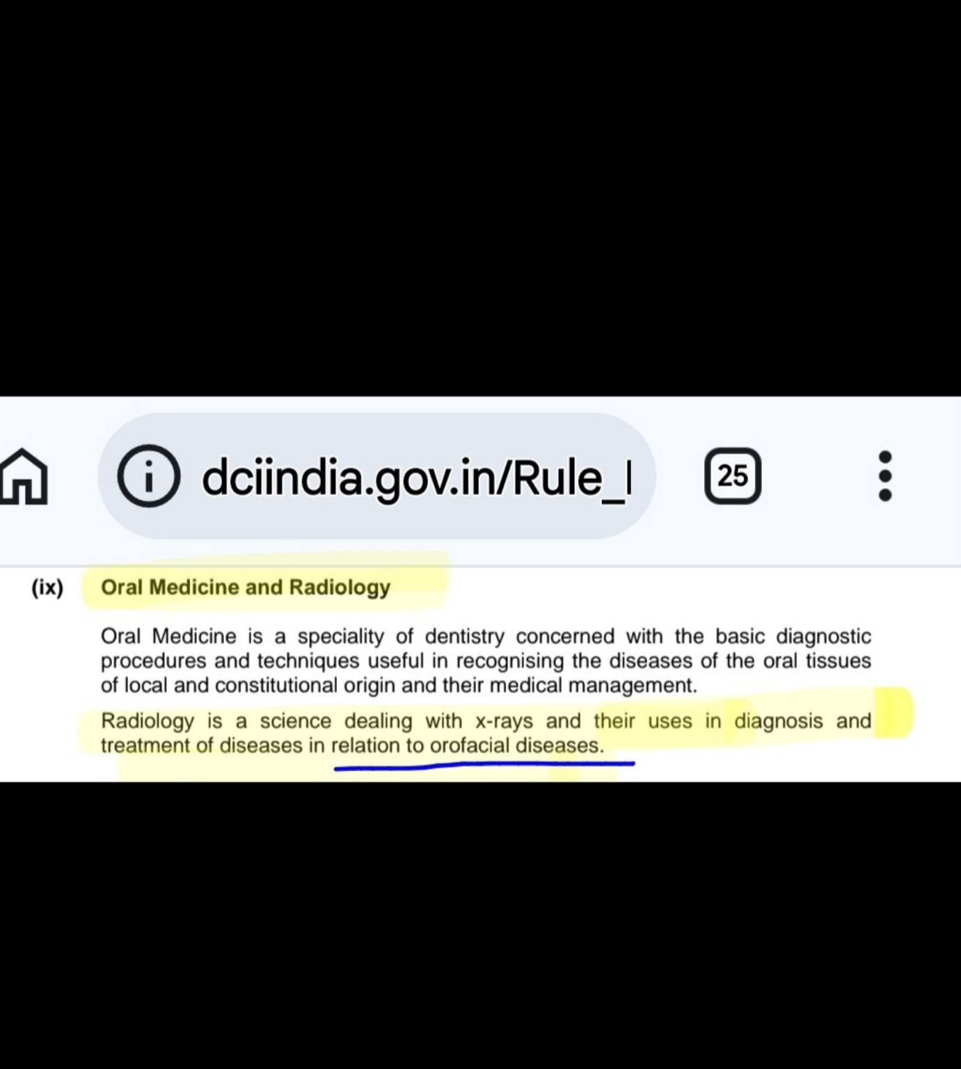
The DCI curriculum describes Oral Medicine as a specialty concerned with basic diagnostic procedures and techniques for recognising diseases of the oral tissues, including diseases of local and constitutional origin, and their medical management. It describes Radiology as the science dealing with X-rays and their use in the diagnosis and treatment of diseases in relation to orofacial diseases. This evidence documents the curriculum’s stated role of radiology in the diagnosis of orofacial diseases.
Original DCI curriculum screenshot – Curriculum evidence (Oral Medicine and Radiology)
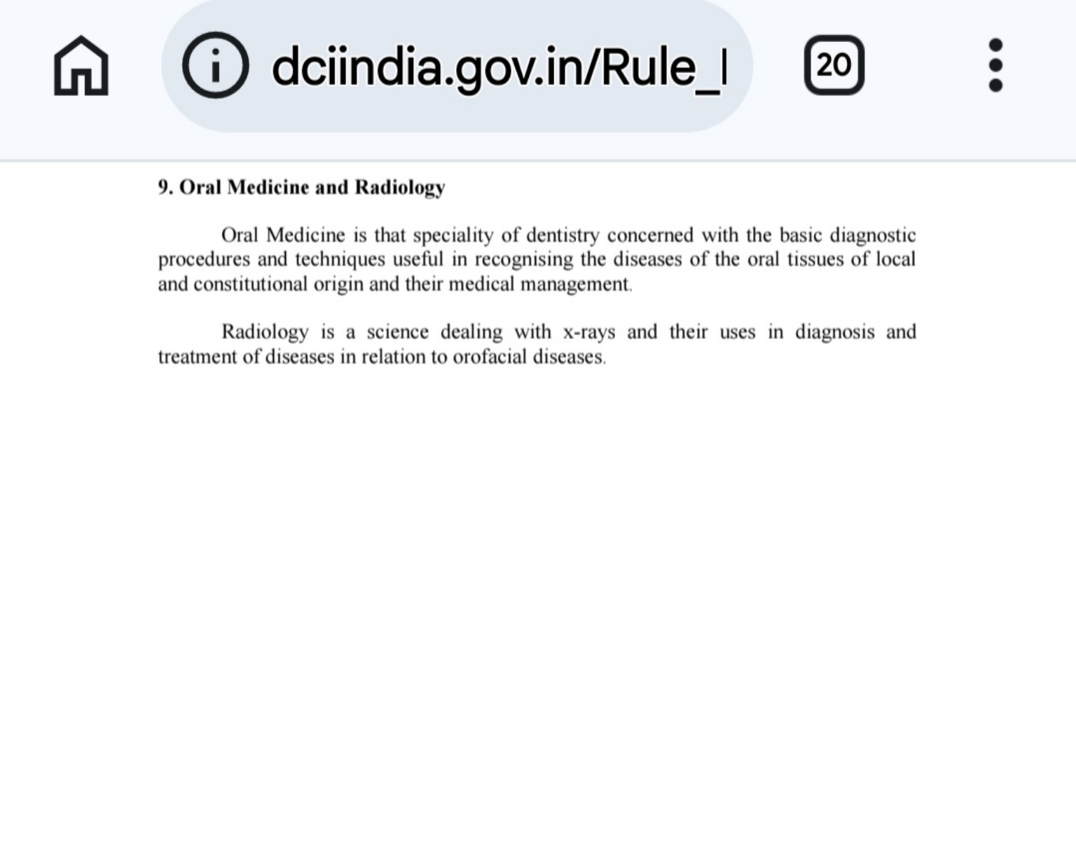
Official source: Dental Council of India – curriculum, Section 9: Oral Medicine and Radiology.
Back to all evidenceCurriculum evidence · Imaging
Dental Curriculum: Radiological Techniques and X-ray Interpretation of the Head and Neck
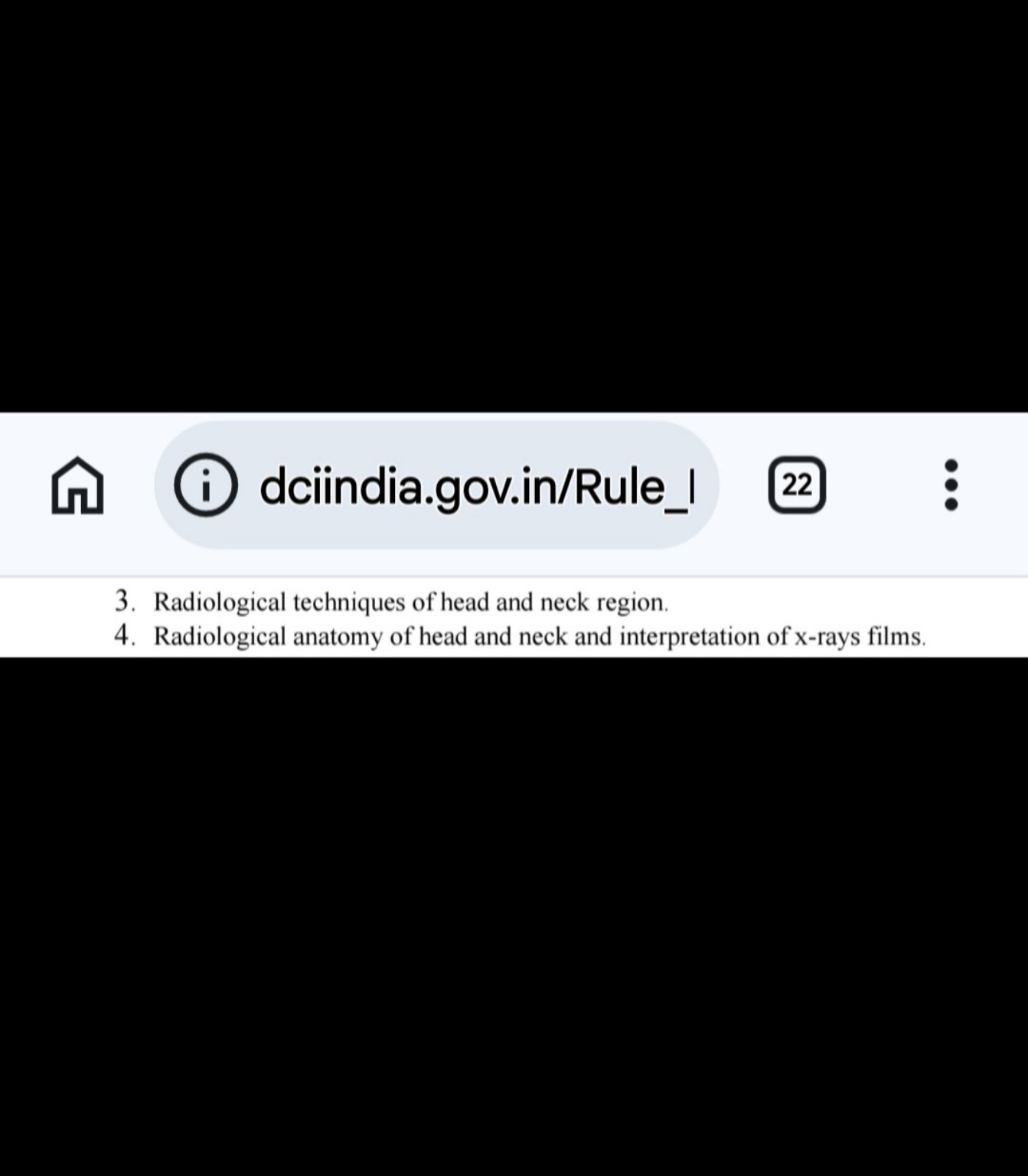
The DCI syllabus excerpt lists radiological techniques of the head and neck region, radiological anatomy of the head and neck, and interpretation of X-ray films.
Original DCI curriculum screenshot – Curriculum evidence
Curriculum evidence · Imaging
Dental Curriculum: Maxillofacial Radiology, Radio Diagnosis and Imaging Techniques
The DCI curriculum excerpt specifically lists Maxillofacial radiology/radio diagnosis and other diagnostic methods and imaging techniques.
Original DCI curriculum screenshot – Curriculum evidence
Curriculum evidence · Imaging
Oral Medicine and Radiology: Radiological Diagnosis of Orofacial Diseases
The DCI curriculum describes Oral Medicine as a specialty concerned with basic diagnostic procedures and techniques for recognising diseases of oral tissues and their medical management. It describes Radiology as a science dealing with X-rays and their uses in diagnosis and treatment of diseases in relation to orofacial diseases.
Original DCI curriculum screenshot – Curriculum evidence
Curriculum evidence · Imaging
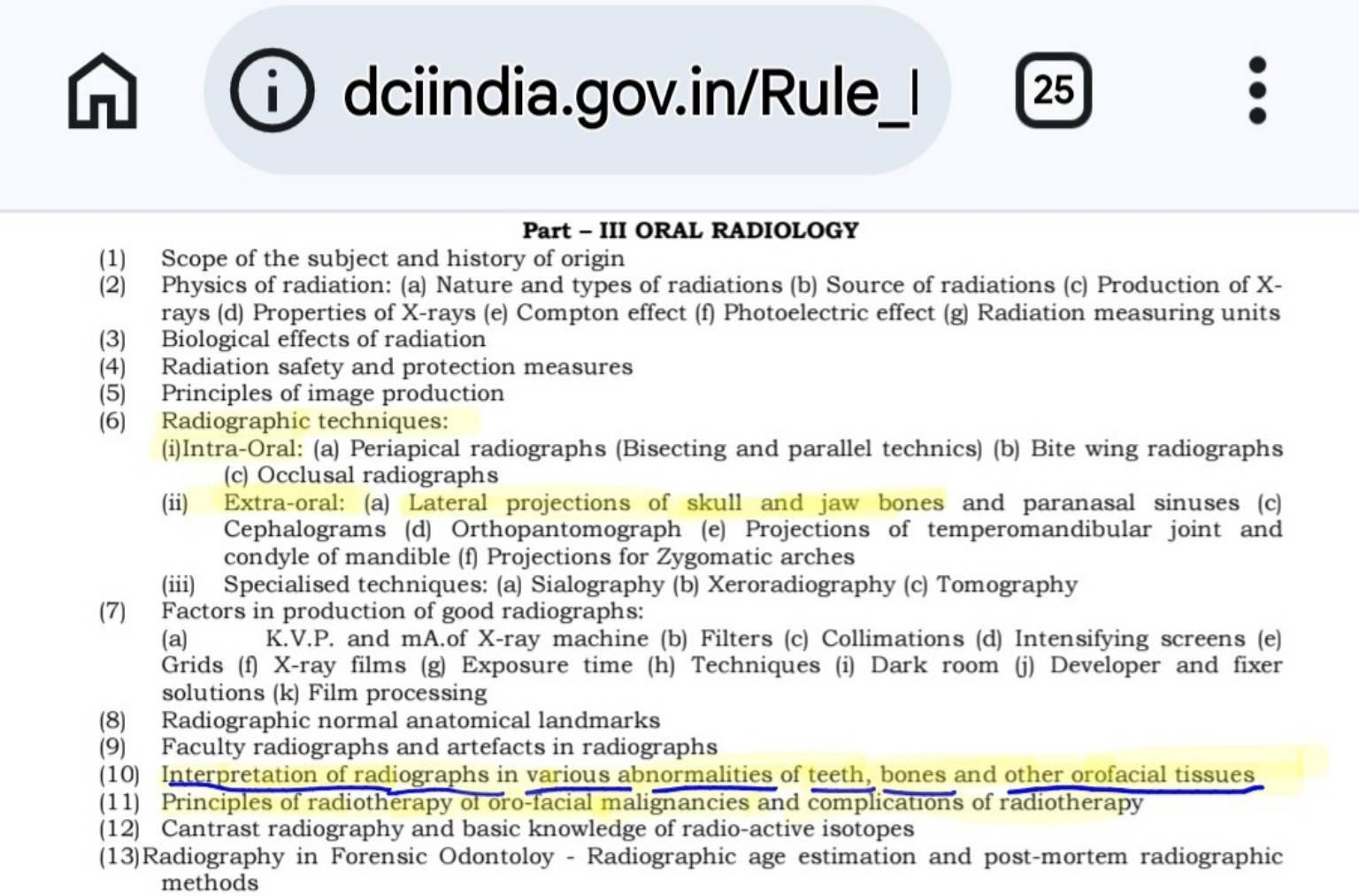
Dental Radiographic Diagnosis and Interpretation of Teeth, Jaw Bones, Skull and Orofacial Tissues
The DCI Oral Radiology syllabus lists intraoral radiographic techniques and extraoral projections of the skull and jaw bones, paranasal sinuses, temporomandibular joint and mandibular condyle, and zygomatic arches. It also specifically includes interpretation of radiographs showing abnormalities of teeth, bones and other orofacial tissues.
Original DCI curriculum screenshot – Curriculum evidence (Oral Radiology)
Understanding curriculum evidence
These original curriculum excerpts illustrate subjects, training objectives or procedures included in the cited dental curriculum. They do not establish that every dentist has the same specialist qualification or is authorised to perform every procedure. A patient’s care should be provided by a clinician whose qualifications, training, competence and applicable regulations match the treatment required.

